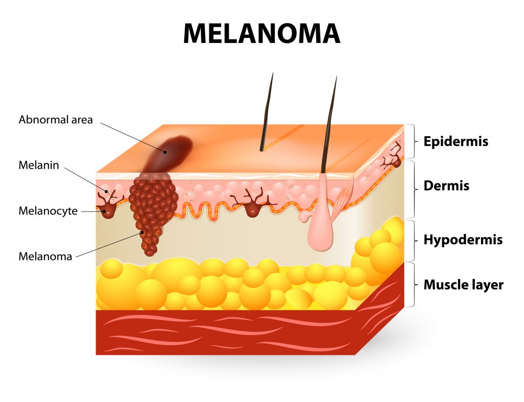
It is a malignant neoplasm…

Melanoma originating from melanocytes, the frequency of which is constantly increasing. It is usually found on the skin. It is the most common malignancy in women aged 25-29 and the second most common after breast cancer in women aged 30-35.</p >

Early diagnosis and treatment often lead to complete healing.</strong >

Aggravating factors are:

  • Light skin
  • Atypical nevi on protected and exposed skin areas
  • History of melanoma
  • Family history of atypical nevi or melanoma
  • History of sunburn
  • Congenital melanocytic nevi

Early symptoms such as an increase in size or change in colour or shape of a nevus as well as itching, tenderness and bleeding should impel you to see your dermatologist immediately.

Melanoma is a malignant tumour originating from melanocytes of the chorio-epidermal boundary. It occurs mainly on the skin but can also be found on the mucous membranes, eyes and meninges. It mainly affects the white race, the average age of diagnosis is 55 years and both sexes have an equal probability of being affected.

Melanoma: what are the risk factors?

  • UV exposure
  • Phenotypic characteristics
  • Phototypes according to Fitzpatrick I and II
  • High number of melanocytic or presence of dysplastic nevi
  • Large congenital nevi
  • History of melanoma
  • Family history of melanoma
  • Inherited mutation in CDK4 and CDKN2A genes
  • Xeroderma pigmentosum

What are the clinical features of melanoma

In the usual clinical types, the biological development of melanoma is distinguished into two phases:

Horizontal Growth Phase: cancer cells spread peripherally or horizontally along the basal layer of the epidermis and papillary dermis, clinically forming a pigmented spot or plaque that expands asymmetrically. This phase is long-term and lasts from months to years.

Vertical Growth Phase: follows the horizontal growth phase and is characterised by the infiltration of the deeper dermis and subcutaneous fat. It presents clinically in the form of a nodule that forms within a short period of time, compared to the previous phase, and in which the cancer cells grow vertically, infiltrating the deeper layers of the skin. This infiltration increases the possibility of metastatic spread of the tumor and worsens the prognosis.

On the basis of histogenetic characteristics, four types of melanoma are distinguished:

  1. Superficial Spreading Melanoma
  2. Nodular Melanoma
  3. Malignant Lenticular Melanoma
  4. Melanoma of the Extremities

What are the symptoms of melanoma?

When a nevus (mole) changes in texture, colour and size, it is a sign that we should contact our dermatologist immediately.

The medical community has designated the “MELANOMA ALPHABET” of the first five letters of the English alphabet (ADCDE) to provide an easy guide to tracking early symptoms of possible melanoma.</ span>

A (Asymmetry)

B (Border)

C (Color)

D (Diameter)

E (Evolving)

What are the factors that contribute to classifying a nevus as dangerous?

Melanoma and Diagnosis

People who belong to an increased risk group should be examined regularly and according to their doctor’s instructions. If there is a genetic predisposition or if there are a lot of moles on the body, they should be systematically monitored and nevus mapping should be performed, an examination in which the medical expert closely monitors any possible change.

What is Nevus Mapping

Digital nevus mapping is the most important means of preventing and controlling skin nevi. The technology in the hands of a dermatologist specialised and experienced in dermoscopy (Dermoscopy Diploma, Medical University of Graz, Austria) enables the early diagnosis of melanoma at a very early stage. For this purpose, a high-resolution dermoscope is used, as well as polarized light, which makes it possible to “photograph” nevi in depth, with very high accuracy.

This examination allows the doctor to monitor and compare the nevi over time, observing in great detail any possible change.

What are the benefits of nevus mapping?

Digital nevus mapping is necessary in the following cases:

  • When there is a history of melanoma
  • When there are multiple nevi
  • When there is a history of burns
  • When there is a weakened immune system

How is the examination of nevi performed at the doctor’s office?

In the first visit, we examine the whole body of the patient, both with the naked eye and with a dermatoscope. In the case that we detect a lesion with melanoma characteristics, we proceed with its removal.

When there is no such lesion, we proceed to the digital imaging of nevi which includes two stages:

  1. The first stage includes the Whole Body Clinical Imaging. This procedure is very important as the technology makes it possible to compare with the clinical photos that will be taken in the following period in order to identify possible new lesions that were not present in previous visits
  2. Dermoscopic Imaging: In this phase we dermatoscopically record all nevi that are larger than two millimeters in diameter and register them topographically with the help of special software. It is this software that allows us to make a comparative assessment and note if the pattern of any nevi differ from others.

This assessment is based on the fact that nevi on the same person have a similar dermatoscopic pattern while melanoma has completely different characteristics that make it stand out.

In case that any damage is detected, our medical expert will decide whether it should be removed immediately or schedule an appointment for it to be rechecked after a short period of time.

Contact us to book an appointment